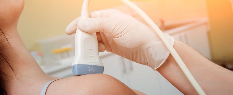
Point of Care Ultrasound continues to assist in the medical practice in a number of ways. Not only does POCUS allow for better imaging and diagnosis from the start, but the ability for imaging to continue to take place during and after a procedure is crucial to patient outcomes. Maverick Medical Education sees application for POCUS in numerous ways and urges all medical practitioners to become familiar with how this technology can benefit patients in all fields. POCUS can help significantly with identifying and treating a fairly common clinical presentation, shoulder dislocation.
Anatomy and Background
According to the American College of Emergency Physicians, “the large range of motion of the shoulder with minimal inferior tendinous support makes it prone to dislocation.” Approximately 50% of all major joint dislocations are shoulder dislocations. Most shoulder dislocations are anterior and caused by larger external force pushing below the glenoid fossa, against the humeral head. Fewer shoulder dislocations are posterior and more difficult to detect in an examination.
Use of POCUS
While standing behind the impacted shoulder, place imaging technology in front of the patient allowing better manipulation of the ultrasound while viewing the screen. Supporting the elbow, the patient should move their arm toward the midline of the body, if possible. Locate the scapular spine and put the probe just under the spine, parallel to it. Detect the humeral head, a round object, which will be deep on the screen rather than lateral with the glenoid fossa in an anterior dislocation. In a posterior dislocation, the humeral head will be very close on the screen. The reduction can take place to reorient the humeral head into correct positioning. After reduction, the position should be corrected and the patient should be able to rotate the shoulder while still holding the limb close to the midline of the body. Additionally, POCUS can be used to confirm everything is in place.
Benefits of POCUS
While plain film radiology can provide some imaging of a patient, the use of POCUS gives a better look into the patient and what needs to be done. Several studies have shown that POCUS is superior in “detecting both anterior and posterior shoulder dislocations, making it another rapidly evolving tool to improve accuracy, decrease error, and improve efficiency.” Further, the portability of POCUS allows for more imaging to take place to ensure the reduction was done properly. If the procedure needs to be repeated, it can be done right away while the patient is still comfortable from sedation, without transporting back to radiology and waiting for plain film x-rays to take place. By eliminating this extra time, patient outcomes are improved with less time in the hospital, saving resources for both the patient and the hospital itself.
To learn more about Point of Care Ultrasound, how to use it, and how it can benefit your community, contact us today. We offer additional courses in pain management techniques. Maverick Medical Education can help you in your practice, today.

